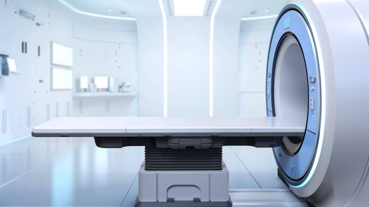
Magnetic resonance imaging (MRI) is a valuable tool for diagnosing many diseases, from liver disease to brain tumors. However, patients must remain completely still during the examination to avoid compromising the quality of the images obtained.
A new prototype device could revolutionize this procedure by detecting patient movements and interrupting the test in real time, improving the experience for patients and technicians.
During an MRI scan, the patient must remain completely still for several minutes in a row. In fact, the slightest movement can generate “Movement artifacts» That blurs the final image. To ensure a clear image, it is necessary to identify any patient movement as soon as it occurs, allowing the scan to be stopped and a new acquisition performed by the technician.
Motion tracking can be achieved using Sensors integrated into the MRI table. Magnetic materials cannot be used because metals interfere with the MRI technique itself.
Particularly appropriate technology for this unique situation, which avoids the use of metallic or magnetic components, is Triboelectric nanogenerator (TENG). The TENG is self-powered using static electricity generated by friction between the polymers.
Li Tao, Zhiye Wu and their colleagues designed a TENG-based sensor that can be integrated into an MRI machine to help prevent motion artifacts. The team created the TENG by placing two layers of plastic films coated with graphite conductive ink around a central layer of silicon. The materials were specially selected so that they would not interfere with the MRI scan.
In tests, when a person turns his head from side to side or lifts it off the table, the sensor detects these movements and sends a signal to the computer. An audible alert will then sound, a pop-up window will appear on the technician's computer screen and the MRI scan will stop.
The researchers say this work could help make MRI scans more efficient and less frustrating for patients and technicians, by producing better images in a single procedure. The prototype device described in ACS Sensors therefore opens new horizons for improving patient diagnosis and monitoring, while improving advances in MRI scans.
Caption: This self-powered sensor (circled in yellow) mounted on the headrest of an MRI machine can help make image collection more efficient by detecting a patient's movements in real time.
Article: “Flexible sensor for real-time monitoring of motion artifacts in MRI” – DOI: 10.1021/acssensors.4c00319


